Fig. 3.
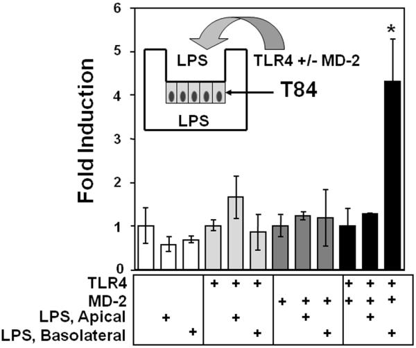
The response to LPS is polarized to the basolateral membrane of T84 cells. T84 were transfected with ELAM-NF-κB-luciferase (0.4 μg) and co-transfected with 0.3 μg of MD-2, TLR4 or both as indicated. Cells were then cultured until TER>2000 Ω-cm2. Lipopolysaccharide 50 ng/ml was added to the apical or basolateral well as indicated. Cells were exposed to LPS for 5 h and lysed for luciferase activity. The data are expressed as fold-induction of relative light units when compared with transfection of the vector control. Only basolateral addition of LPS in cells expressing both TLR4 and MD-2 resulted in NF-κB activation. These data are one representative experiment of three independent experiments performed in triplicate and error bars indicate standard deviation.

