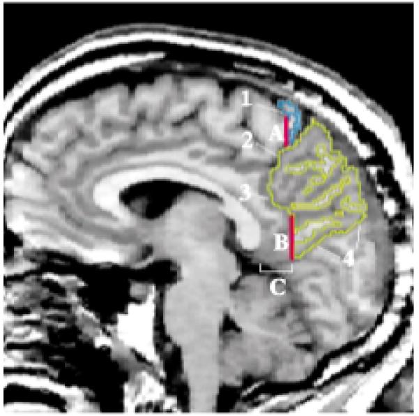
FIGURE 6. Regions of the Parietal Lobea.

a Image depicts a sagittal MRI slice approximately 5 mm lateral to the midsagittal slice, with the superior parietal gyrus traced in yellow and the postcentral gyrus in blue.

a Image depicts a sagittal MRI slice approximately 5 mm lateral to the midsagittal slice, with the superior parietal gyrus traced in yellow and the postcentral gyrus in blue.