
FIGURE 8. Boundaries Defining Plane B (from figure 7) of the Cortexa.
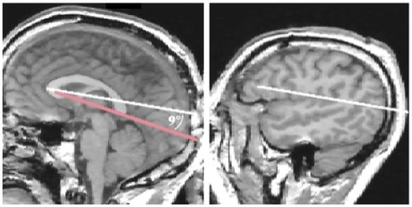
a The left side of the figure shows a midsagittal MRI slice with the reference line from figure 5 in red. The white line was drawn from the most anterior to the most posterior point of the corpus callosum, which formed a 9° angle with the reference line. The right side of the figure depicts the second line that defined the boundary of plane B, which was drawn on a lateral (sagittal) MRI slice with the same x, y coordinates as the red reference line.