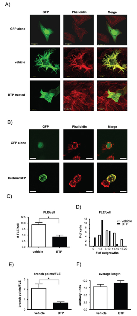
Figure 4. BTP inhibits drebrin function.
A) CHO cells transfected with either GFP (top panel) or GFP-drebrin (middle and bottom panels). Cells were treated with DMSO (vehicle, middle panel) or BTP (bottom panel) prior to staining for F-actin (red) and visualization using confocal laser scanning microscopy. One of 3 similar experiments. B) Jurkat T cells were transfected with either GFP (top panel) or GFP-drebrin (bottom panel). Cells were stained for F-actin (red) and visualized as in (A). One of at least 5 similar experiments. White bar indicate 20 µm. C) Branched cell extensions caused by drebrin over-expression (filopodia-like extensions, FLE) were counted on each cell in DMSO and BTP treated cells and expressed as average FLE per cell, D) the number of cells having a given range of FLEs per cell, E) the average number of branch points per FLE, and F) the average length of each FLE. For all measurements n=25 cells, *p<0.05. One of 3 similar experiments.

