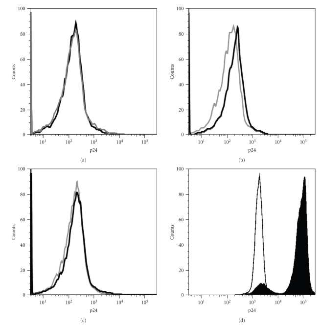
Figure 3.
CD8+ T having acquired CD4 bind HIV particles. (a) Expression of p24 on CD8+ T cells having captured (open black histogram) or not (closed grey histogram) CD4 in a previous coculture (as shown in Figure 2 but target cells are HEK-FcR stably transfected with human CD4) and incubated with uninfected MOLT-4 cells. Shown are overlaps of p24 staining on gated CD8+ T cells. (b) As in (a) except that infected MOLT-4 cells were used in the coculture with CD8+ T cells. (c) As in (b) except that the Leu3a neutralizing anti-CD4 mAb was present during the coculture. (d) Shown are overlap of p24 staining on uninfected MOLT-4 cells (as in condition shown in (a), open black histograms) and on infected MOLT-4 cells (as in conditions shown in (b) and (c), closed black histograms). Note that MOLT-4 cells were analyzed from the coculture with T cells and with the very same settings used for the analysis of T cells, which explains the high autofluorescence of MOLT-4 cells, which is related to their larger size. Similar results were obtained in a second independent experiment.

