
Fig. 2.
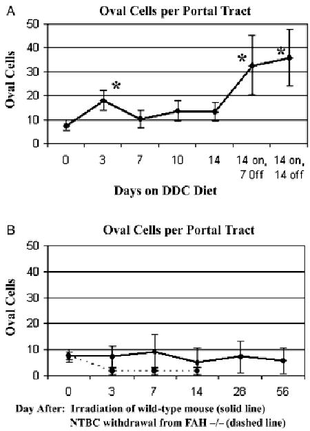
Time course of oval cell proliferation (mean ± SE, *P < 0.05 by student's t-test). Normal oval cell density is indicated as a dotted horizontal line. (A) DDC diet – there was a modest increase in oval cell density by day 3, becoming more marked during the recovery period 7–14 days after stopping DDC (stars). (B) Liver irradiation and FAH-null mouse after NTBC withdrawal. There were no significant deviations from normal in oval cell density after focal liver irradiation; FAH-null mouse shows diminished proximal biliary structures and no oval cell proliferation. DDC, 3,5-diethoxycarbonyl-1,4-dihydrocollidine; FAH, fumaryl acetoacetate hydroxylase.