
Figure 1. The pool of quiescent Pax7 satellite cells returns to homeostasis during adult muscle regeneration.
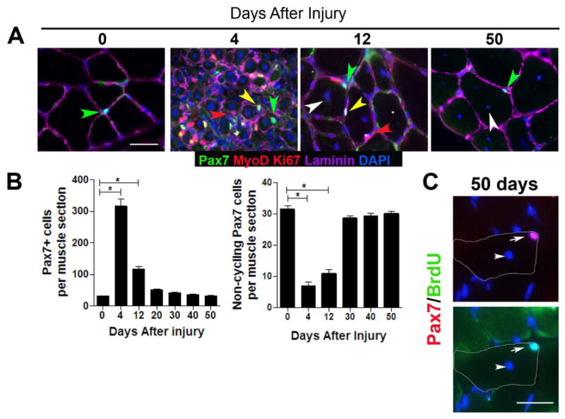
(A) Transverse sections of uninjured (0 days after injury) and regenerating (4, 12, and 50 days after injury) TA muscle contain quiescent (MyoD-, Ki67-) (green arrowhead) and cycling (MyoD+, Ki67+; yellow arrowhead) Pax7+ cells in sublaminar location (laminin+; magenta). Differentiating myogenic cells (red arrowhead) could be observed in regenerating muscle underneath the basal lamina. (B) Total number of Pax7+ cells (left) and quiescent (Ki67-; MyoD-) Pax7+ cells (right) per muscle section from 4-6 mice per time point (mean ± sem; p<0.05). (C) BrdU+/Pax7+ cells in transverse sections from fully regenerated muscle from BrdU-treated mice. Top panel: Pax7 (red) and DAPI (blue) staining, Bottom panel: BrdU (green) and DAPI staining. Regenerated muscle fibers are distinguished with DAPI+ central nuclei (white arrowhead). The contour of muscle fiber is denoted by white line. Scale bar; 40 μm (A), 80 μm (C).