
Figure 2. Pax7-derived satellite cells are capable of self-renewal and differentiation.
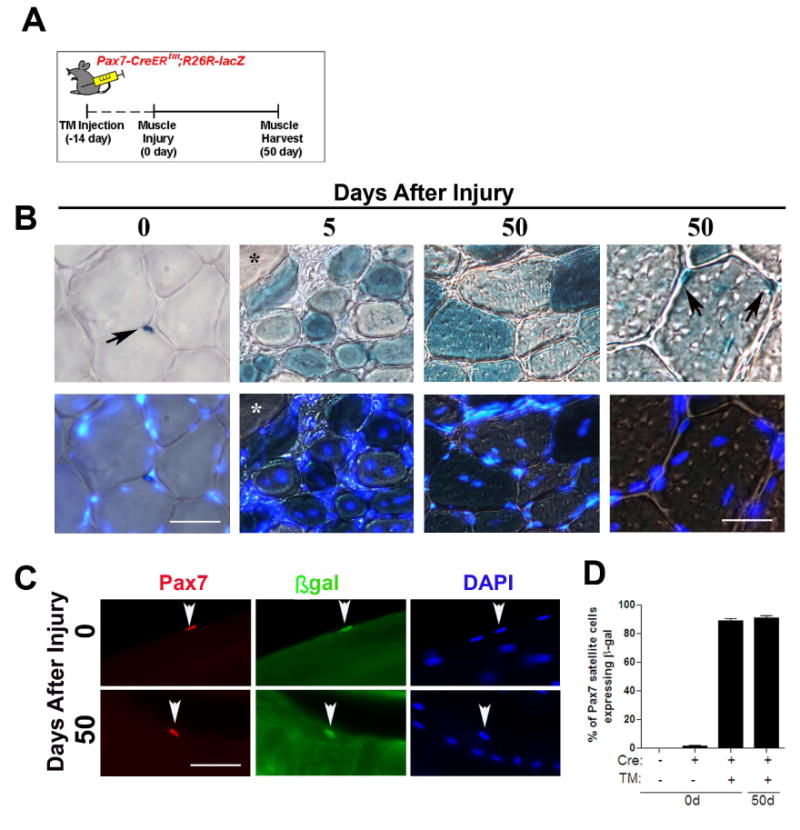
(A) The cartoon depicts the tamoxifen (TM) injection scheme for lineage tracing muscle satellite cells. Pax7+ satellite cells in the Pax7-CreERtm;R26R-lacZ mouse were permanently labeled by IP administration of TM 14 days prior to muscle injury. (B) Transverse sections were collected from uninjured and regenerating muscle and stained with X-gal. Five days after injury non centrally-nucleated fibers in regenerating muscle had no detectable X-gal reactivity (*). After 50 days of regeneration, X-gal+ mononucleated cells were observed in the satellite cell position of regenerated muscle fibers (black arrows). (C) Single fibers from uninjured and regenerated Pax7-CreERtm;R26R-lacZ muscle stained with Pax7 (red), β-gal (green) and DAPI (blue). Regenerated muscle fibers are characterized by DAPI+ ‘central-myonuclear chains’. (D) The percentage of β-gal+/Pax7+ satellite cells expressing on single fibers isolated from uninjured and regenerated muscle fibers in the presence (+) or absence (-) of Cre or TM. Data is presented as mean ±sem. n=4-6 mice. (p<0.05). Scale bars in (B) are 60 μm (left) and 30 μm (right) and 40 μm in (C).