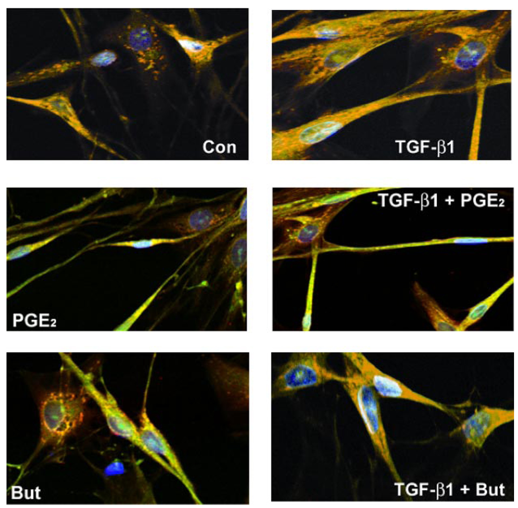
Fig. 10.
PGE2 alters cell morphology and inhibits the formation of TGF-β1-induced focal adhesions in adult lung fibroblasts. Normal adult lung fibroblasts were serum-starved for 48 h before culture with serum-free media alone, TGF-β1 alone (2 ng/ml), PGE2 alone (100 nM), butaprost alone (5 µM), or TGF-β1 in combination with PGE2 or butaprost. Cells were then fixed and stained with FITC-phalloidin (green), paxillin (red), and DAPI staining of nuclei (blue). Cells were then analyzed using laser-scanning confocal microscopy with appropriate wavelengths using a ×60 water immersion objective. Merged images are shown, and focal adhesions appear as orange/yellow. Z-stack analysis confirmed the colocalization of the FITC and indocarbocyanine (Cy3) staining.

