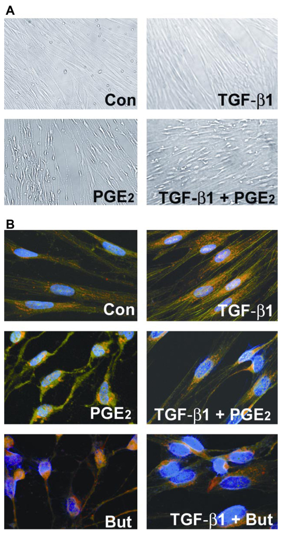
Fig. 3.
PGE2 induces sudden and sustained morphological changes. A: IMR-90 cells were growth-arrested at 85% confluency for 48 h. Medium was replaced with serum-free media or that containing TGF-β1 (2 ng/ml) or PGE2 (10 nM) or both TGF-β1 and PGE2. Cells were photographed after 5 min of treatment. Control (Con) cells show characteristic flattened spindle shape. TGF-β1-treated cells show spindle shape with a more 3-dimensional appearance. PGE2-treated cells are no longer spindle shaped and appear shrunken longitudinally with decrease in area of surface fibrillar and focal adhesion. The appearance of the cells treated with both TGF-β1 and PGE2 is intermediate between those treated with TGF-β1 alone and those treated with PGE2 alone. B: PGE2 alters cytoskeletal structure and focal adhesions in human lung fibroblasts. Shown are FITC-phalloidin staining (green), immunocytochemistry with paxillin (red), and 4,6-diamidino-2-phenylindole (DAPI) staining of nuclei (blue). IMR-90 cells were serum-deprived for 48 h. Medium was replaced with serum-free media or that containing TGF-β1, PGE2, butaprost (But), or combinations of both TGF-β1 and PGE2 or butaprost. After 24 h, cells were washed, fixed, and stained. Cells were then analyzed using laser-scanning confocal microscopy with appropriate wavelengths using a ×60 water immersion objective. Merged images are shown, and focal adhesions appear as yellow/orange.

