Fig. 4.
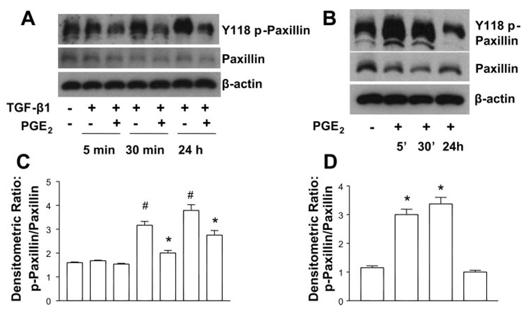
PGE2 inhibits the TGF-β1-stimulated phosphorylation of paxillin. A: quiescent human lung fibroblasts (IMR-90) were treated with TGF-β1 (2 ng/ml), or TGF-β1 (2 ng/ml) and PGE2 (10 nM), under serum-free conditions, and cell lysates were prepared at the times indicated. B: quiescent human lung fibroblasts (IMR-90) were treated with PGE2 (10 nM) under serum-free conditions, and cell lysates were prepared at the times indicated. Cell lysates were subjected to SDS-PAGE followed by immunoblotting with antibodies against p-paxillin. Blots probed with antibody against p-paxillin were stripped and blotted with antibodies against paxillin and β-actin sequentially. C: densitometry data for n = 3 experiments as in A. #P < 0.05 for TGF-β1-treated samples compared with untreated control; *P < 0.05 for TGF-β1 + PGE2-treated samples compared with TGF-β1 treatment alone. D: densitometry data for n = 3 experiments as in B. *P < 0.05 compared with untreated control.

