
Figure 4. Subcellular localization of the V2 splice variants.
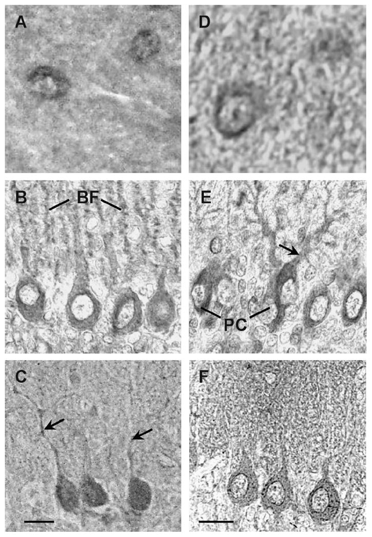
High magnification of cerebellar sections stained with the AS382 (A–C) or with ASC-3 (D–F). Immunopositive neurons in the molecular layer at P30 (A, D). At P15 thesomata of Purkinje cell (PC) and Bergmann fibers (BF) are immunostained (B); at P5 the somata of Purkinje cell and growing dendrites are immunostained (E). At P30 thesomata of Purkinje cells (C–F) and dendrites are immunopositive for the V2a (C, arrows). Scale bars, 20 μm.