
Figure 7. The V2b splice variant down-regulated the surface expression of V2a receptor.
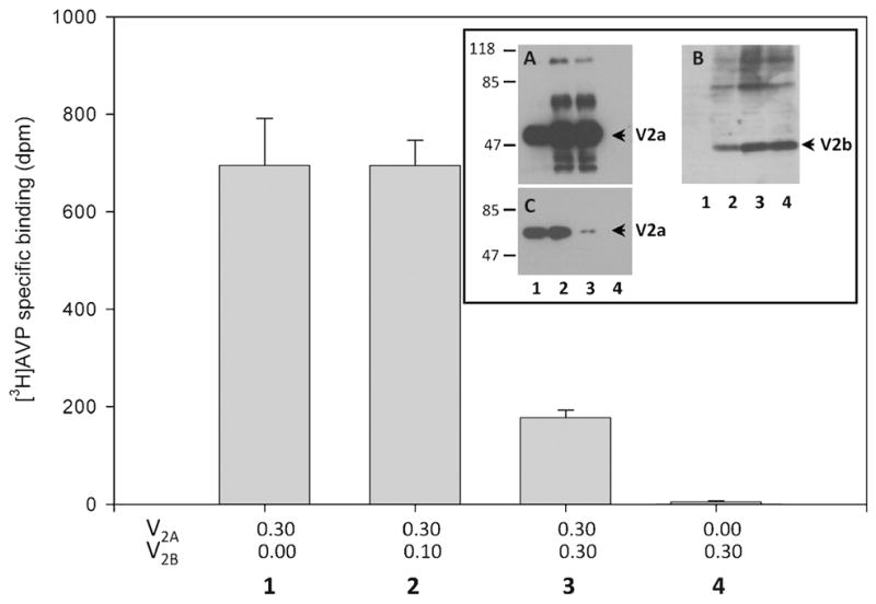
CHO K1 cells transfected with increasing amounts of cDNA encoding V2b (μg) and a constant amount of cDNA encoding V2a (bars/lanes 1–3). Cells transfected with V2b alone did not show [3H] AVP specific binding (bar 4). The inset shows Western blots of transfected cell extracts using anti-GFP to detect V2a tagged with GFP (A) and anti-HA to detect V2b tagged with HA (B). The V2a protein expression does not significantly change in cells transfected with increasing amounts of V2b cDNA (A); the expression of V2b increased as the amount of V2b cDNA employed for transfections (B). Western blot analysis of biotin-labeled proteins precipitated with neutravidin agarose beads and stained with anti-GFP antibodies (C). The numbers (1–4) correspond to the bar and lanes (inset) and indicate the amount of cDNAs (μg) employed in the transfections. Data are the average of three experiments.