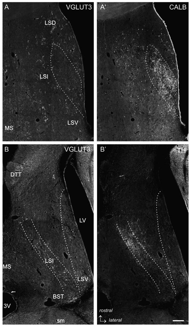
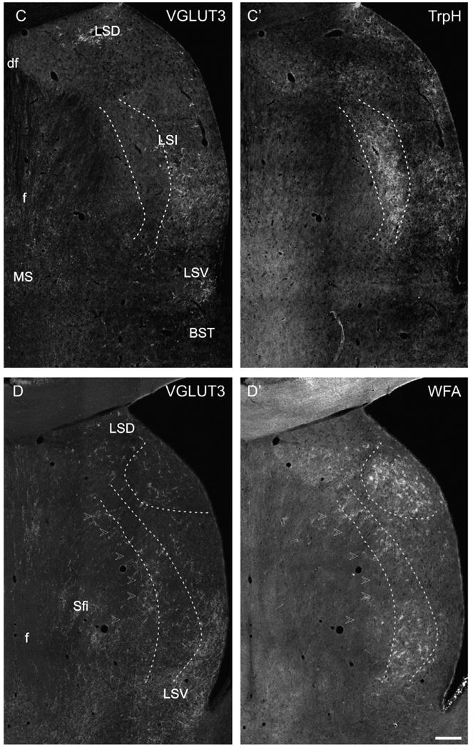
Fig. 3.


Low power photomicrographs of VGLUT3-immunofluorescence labeling with different markers. (A/A′) VGLUT3-CALB double labeling, intermediate level of the LS. The dashed lines delineate the bulk of CALB-ir projection neurons of the LS. In this region, only a few VGLUT3-ir PBs are found, whereas in the medial LSI, LSD and LSV loosely scattered CALB-ir cells intermingle with VGLUT3-ir PBs. (B/B′) VLGUT3-TH double labeling in a horizontal section of the LS (Bregma −5.82 mm according to Paxinos and Watson, 1998). It is appreciable that the TH-ir fiber network strongly overlaps with the band of VGLUT3-ir in the mediorostral but not in the laterocaudal LSI. (C/C′) VGLUT3-TrpH double labeling in a caudal section of the LS illustrating a dense cluster of TrpH-ir fibers and baskets in the medial LSI (dashed lines) which only exceptional overlaps with VGLUT3-ir PBs. In contrast, the TrpH-ir fibers in the lateral and subventricular LSI intermingle with VGLUT3-ir. (D/D′) VGLUT3–WFA double labeling. In the caudal LS, an onion skin-like stripe that overlaps with the VGLUT3-ir band (arrowheads) of less dense WFA-staining is present. Additionally, dense WFA-binding was visible in the LSD (dashed lines). Scale bar (valid for A–D) = 500 μm.
