
Fig. 1. Regenerated cardiomyocytes are derived from differentiated cardiomyocytes.
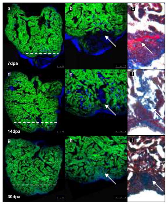
Cardiomyocytes in transgenic zebrafish (tg-cmlc2a-Cre-Ert2: tg-cmlc2a-LnL-GFP) were genetically labelled at 48 hpf by inducing Cre activity with tamoxifen. These embryos were then grown to adulthood (3months/sexually mature) at which point the heart was amputated and allowed to regenerate for 7 (a, b, c), 14 (d, e, f) or 30 days (g, h, i). Dashed white line represents the plane of amputation. At 7 dpa (a, b) relatively little regeneration has occurred. Trichromic staining indicates a fibrin clot has formed adjacent to the wound (c). By 14 dpa, GFPpos cardiomyocytes have regenerated a substantial amount of new cardiac tissue (d, e) and the fibrin clot is reduced in size (f). At 30 dpa, heart regeneration is virtually complete (g, h) and all of the regenerated tissue is comprised of GFPpos cardiomyocytes. The clot has been replaced by a small scar (h). Scale bars represent 100 μm in a, d, g, and 75 μm in b, e, h.