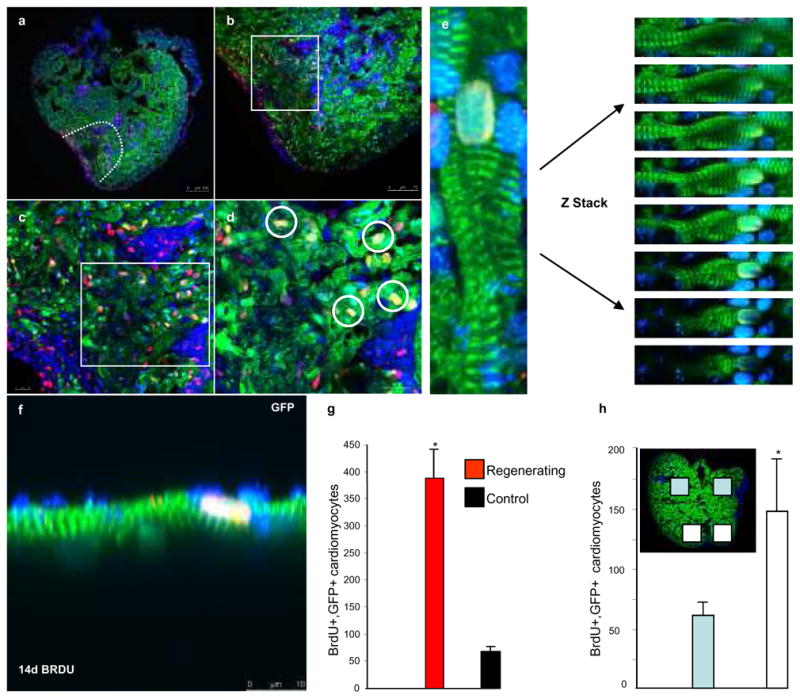
Fig. 2. Differentiated cardiomyocytes re-enter the cell cycle.

Transgenic zebrafish (tg-cmlc2a-Cre-Ert2: tg-cmlc2a-LnL-GFP) genetically labelled at 48 hpf and grown to adulthood were amputated then treated with BrdU at 7 dpa, hearts were then isolated and processed at 14 dpa (a–f). Green= GFPpos cardiomyocytes; Red=BrdUpos cells; Blue= DAPI; Yellow= BrdUpos/GFPpos cardiomyocytes (white rings in d).(g) Indicates the average number of BrdUpos/GFPpos cardiomyocytes/section +/− SEM, t-test * p<0.01; amputated n=17 sections from 7 different animals, control n=9 sections from 3 different animals. (h and inset) Indicates the distribution of BrdUpos/GFPpos cardiomyocytes (n=5 sections from 5 different animals). Scale bars represent 100 μm in a, 75 μm in b, and 10 μm in c, d, f.
