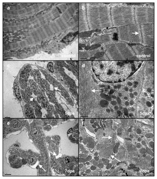
Fig. 3. Cardiomyocytes dedifferentiate resulting in the disassembly of sarcomeric structure and detachment.
Electron microscopy of a control heart (a, b), a 5-dpa regenerating heart (c, d) and a 7-dpa regenerating heart (e, f). Cardiomyocytes in unamputated control samples show a tightly organised sarcomeric structure (a), at higher magnification (b) the Z-lines are clearly visible (white arrow). At 5 dpa many of the cardiomyocytes display a disorganised sarcomeric structure (c) along with the appearance of intercellular spaces (white arrows). Closer examination reveals a loss of Z-lines (d, white arrow). At 7 dpa there is a similar loss of structure and appearance of intercellular spaces (e white arrows). At higher magnification (f) myosin fibres are visible (arrows) however both longitudinal (upper arrow) and transverse (lower arrow) fibres are present within the same cardiomyocyte indicating disorganised sarcomeric structure. Scale bars represent 0.5 μm in a, b, d, and 2 μm in c, e, f.

