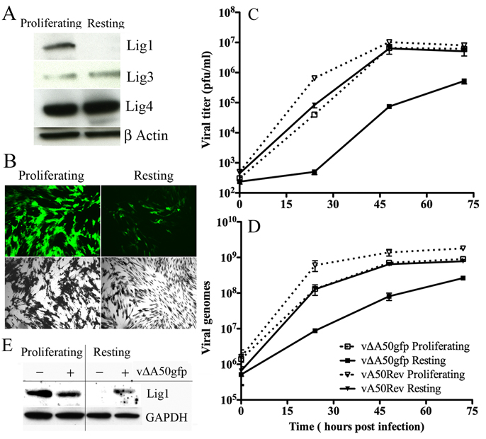
Figure 3. Analysis of Cellular Ligases and Replication of vΔA50gfp Ligase Deletion Mutant in Resting and Proliferating HFF.
(A) Cellular ligases in resting and proliferating cells. HFF were maintained in medium containing 0.2% FBS for four days to induce the resting state or were passaged in medium containing 10% FBS to allow proliferation. The cells were lysed and the proteins analyzed by Western blotting with antibodies to Lig1, 3, or 4 or to β-actin and detected by chemiluminescence. (B) VACV late gene expression. Resting and proliferating HFF were infected with 0.02 PFU per cell of vΔA50gfp. After 48 h, the cells were visualized by fluorescence microscopy (green) and after 72 h by light microscopy after crystal violet staining (black and white). (C, D) Virus replication and DNA synthesis. Resting and proliferating HFF were infected with 0.02 PFU per cell of vΔA50gfp or vA50Rev. At indicated times, virus titers were determined by plaque assay or viral DNA was quantified by real-time PCR. Experiments were in duplicate and bars indicate standard error. (E) Induction of Lig1 in resting cells by VACV. Lig1 and GAPDH were analyzed by Western blotting of extracts from resting and proliferating HFF that were uninfected or infected for 48 h.

