Fig. 4.
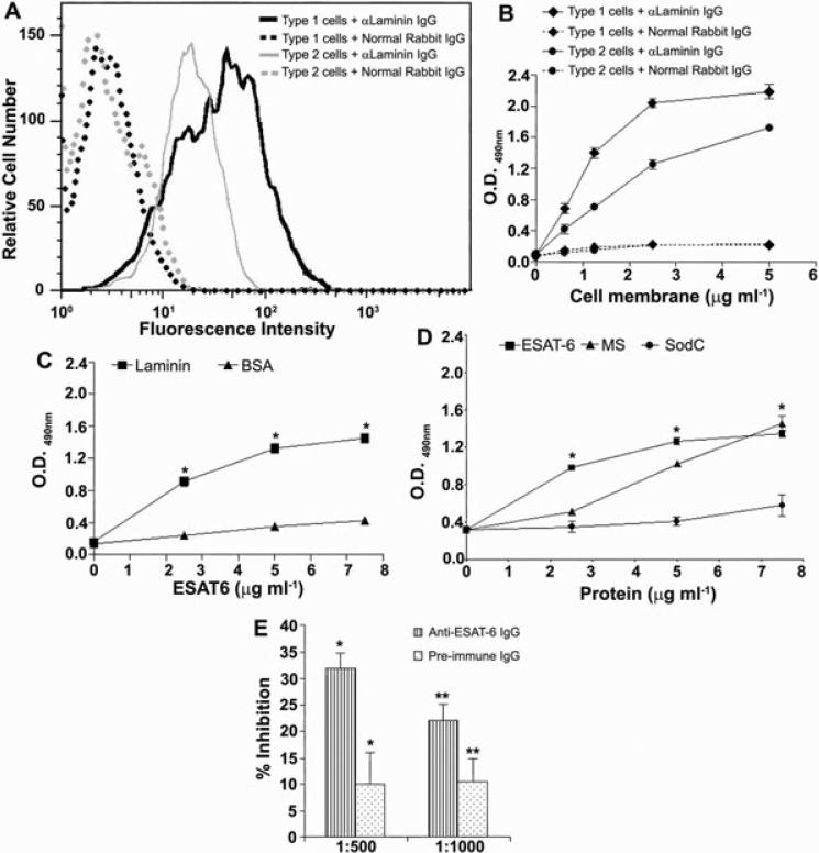
Presence of laminin on the surface of type 1 and type 2 pneumocytes.
A. Cells were exposed to anti-laminin antibodies or normal rabbit antibodies followed by FITC-conjugated anti-rabbit antibodies. Type 1 pneumocytes have more laminin/cell than type 2 pneumocytes. Cells were exposed to each antibody in triplicates and the experiment was done two times.
B. Detection of laminin in membrane preparations of cells. Different concentrations of the membrane preparations were tested in triplicates and the experiment was performed twice with similar results. The OD values (mean ± SD) from one representative experiment are plotted.
C. ESAT6 binds to purified laminin: increasing concentrations of ESAT6 were added to wells coated with laminin or BSA at 1 μg ml–1 and the binding of ESAT6 detected with anti-ESAT6 IgG. Each condition was tested in triplicate and the experiment was performed twice with similar results. Values (mean ± SD) from one representative experiment are plotted. As compared with BSA the binding of ESAT6 to laminin was significantly higher at all concentrations tested (*P < 0.0001).
D. Specific binding of ESAT6 to laminin: various concentrations of His-tagged M. tb proteins (ESAT6, MS and SodC) were incubated with laminin (1 μg ml–1) coated wells and binding of M. tb proteins was detected with anti-His mAbs. Compared with SodC, the binding of ESAT6 to laminin was significantly higher at all concentrations tested (*P < 0.0001).
E. Inhibition of binding of ESAT6 to laminin by anti-ESAT6 IgG. ESAT6 preincubated with anti-ESAT6 IgG (striped bars) or pre-immune IgG (dotted bars) was added to laminin-coated wells (in triplicates) and the laminin-bound ESAT6 was detected by anti-His IgG. Per cent inhibition of binding of ESAT6 to laminin from one representative experiment is plotted. Inhibition of ESAT6–laminin interaction by anti-ESAT6 IgG was significantly higher compared with inhibition by pre-immune IgG at both dilutions tested (*P = 0.009 at 1:500 and **P = 0.037 at 1:1000).

