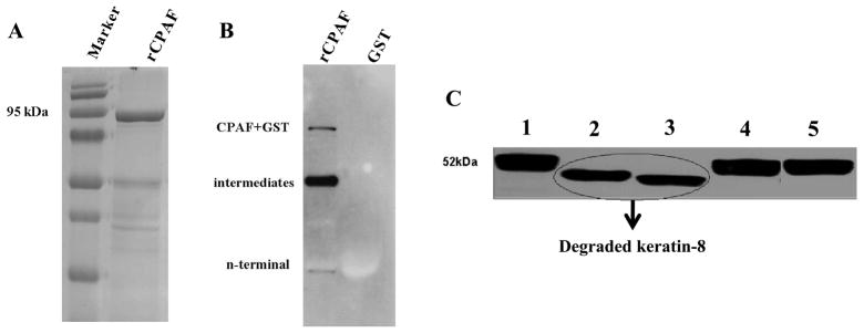
Figure 1. Molecular size and enzymatic activity of rCPAF.
(A) GST-rCPAF was purified using glutathione Sepharose 4B and loaded in a 10% SDS-polyacrylamide gel and stained with coomassie blue. (B) GST-rCPAF and GST alone were probed with mouse anti-CPAF c-terminus monoclonal antibody. (C) Purified active or inactive rCPAF from C. muridarum, or S100 fraction from HeLa cells infected with C. trachomatis (L2S100), or GST were incubated with cytosolic extract (CE) from the HeLa cells at 37°C for 3 hr. The full length and degraded keratin-8 fragments were detected using mouse anti-human keratin-8 primary antibody followed by goat anti-mouse secondary antibody and developed using an ECL reagent. Lanes 1: CE only, 2: CE+native CPAF, 3: CE+active rCPAF, 4: CE+denatured rCPAF, and 5: CE+GST.

