
Figure 4.
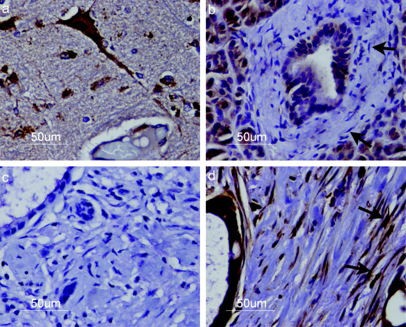
Immunohistochemical staining of Smo protein. Cells were stained with an anti-SMO antibody and an HRP-labeled secondary antibody (brown) then counterstained with hematoxylin and eosin. Staining revealed (a) strong Smo expression in Purkinje neurons of cerebellar tissue; (b) lack of Smo expression in normal pancreatic stromal fibroblasts (arrows) (c) no Smo staining in a primary pancreatic cancer section incubated with secondary antibody alone; and (d) strong SMO expression in pancreatic cancer associated stromal fibroblasts (arrows). Smo expression is also detected in pancreatic cancer cells. Representative samples from each type of tissue are shown. Magnification x40.