
Figure 4. Effects of GGI-58 isoforms on intracellular TAG content and lipid droplet degradation.
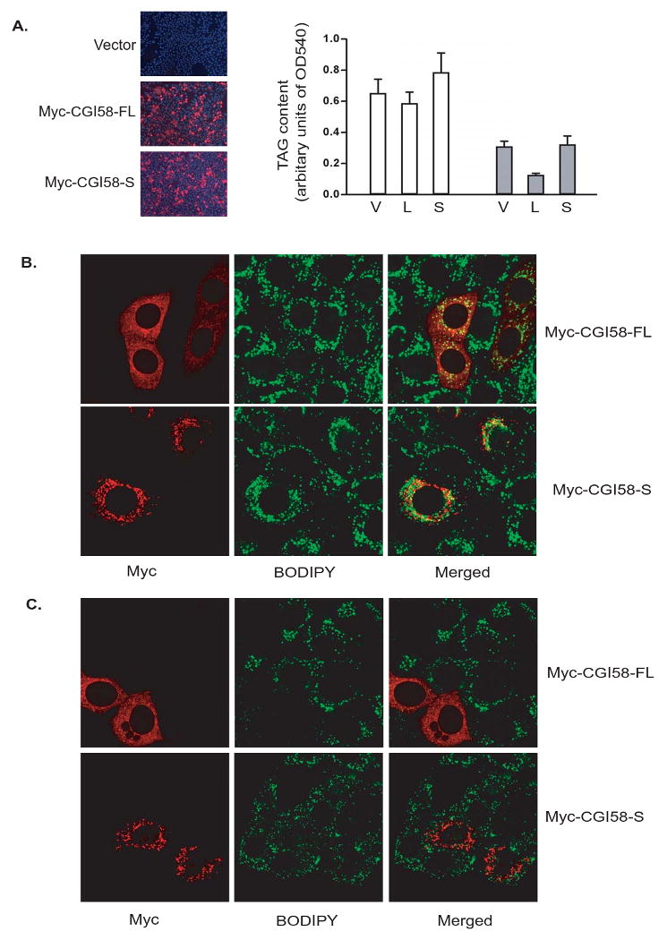
(A) Left panel: HeLa cells were transfected with vector alone (V), Myc-CGI-58FL (L) or Myc-CGI-58S (S). Immunofluorescence staining with anti-Myc antibody (red) was performed to reveal the transfected cells. Nuclei were co-stained with DAPI fluorescence dye (blue). Right panel: Following transfection, cells were incubated under normal growth conditions with 400 μM of oleic acid complexed to albumin for 24 h (white bars). A separate set of cells were incubated in serum- and glucose-free medium for 4h following 24h of incubation with 400 μM of oleic acid complexed to albumin (grey bars). The TAG content was determined as described in Materials and Methods. Data are shown as mean ± SD and represent three independent experiments. (B) HeLa cells transfected with Myc-CGI-58FL or Myc-CGI-58S were mixed with untransfected cells followed by treatment with oleic acid for 24h. Immunofluorescence staining with Myc antibody (red) was performed, and lipid droplets were co-stained with BODIPY 493/503 fluorescence dye (green). (C) Same experiments as in (B) except that cells were incubated in serum- and glucose-free medium for 4h following 24h of oleic acid treatment.