
Figure 5.
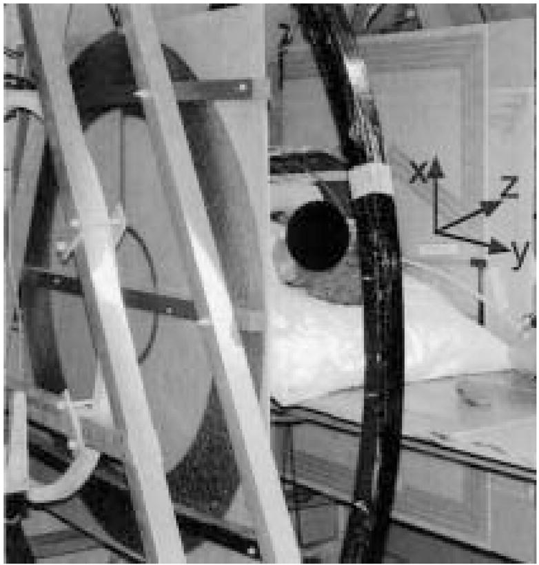
The planar gradient sets mounted on the aluminum support frame of the open-access human MRI B0 coil system. The x and y gradients are placed just inside of the z gradients, relative to the subject (seen in supine position). As such the z-axis gradient is visible on the subject’s left, and the x-axis gradient is seen to his right.