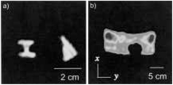
Figure 7.
a) Images of 3He laser-polarization cells, obtained at 4.1 mT with the very-low-field, open-access, human MRI system, using a gradient echo sequence with the following parameters: 132 kHz, 50 cm acquired FOV, 128 × 128 pixels, TE = 10 ms, TR = 500 ms, and two averages. b) 1H image of a large tub containing water, obtained at 3.0 mT with the 3He very-low-field MRI system. A spherical plastic rod was placed across the bottom of the tub to provide image structure. A spin echo sequence was used with the following acquisition parameters: 127 kHz, 125 cm acquired FOV, 128 × 128 pixels, TE = 36 ms, TR = 2.4 s, and 48 averages.

