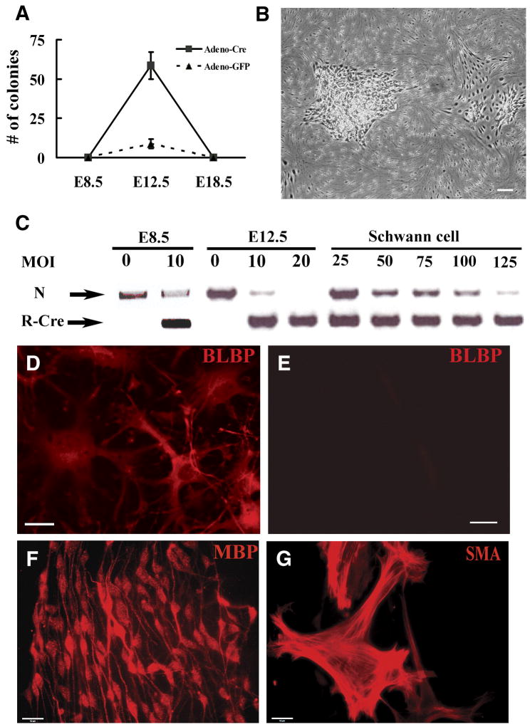
Figure 1. Acute loss of Nf1 in dorsal root ganglion cells at E12.5+1 results in colony formation in vitro.
(A). Quantification of colonies resulting from acute loss of Nf1 in E8.5 neural tube-derived crest cells, E12.5 dorsal root ganglion, (DRG) cells or differentiated Schwann cells (denoted E18.5) derived from Nf1 flox/flox mice cells. A few colonies were observed in the E12.5 DRG cells infected by adeno-GFP, but could not be expanded. (Mean ± SD). (B). Phase micrograph of expandable adherent colonies resulting from E12.5 DRG cells infected by adeno-Cre. (C). Adeno-Cre mediated recombination confirmed by PCR. N: Nf1 flox allele, R-Cre: recombined Nf1 allele. MOI = multiplicity of infection. Cells replated from colonies after E12.5 DRG-derived recombination (D) or adeno-GFP (E) immunostained with anti-Blbp and staining detected with a fluorescently tagged anti-rabbit antibody. (F, G). Differentiation of E12.5 DRG-derived colony-forming cells in vitro. E12.5 DRG-derived colonies were replated and cells cultured under gliogenic and smooth muscle/myofibroblast generating culture conditions for 12 days, immunostained with anti-MBP (F) or anti-SMA (G) antibodies and staining detected with appropriate second antibodies. Bars, B=50μm, D, E=26μm, F, G =15μm.

