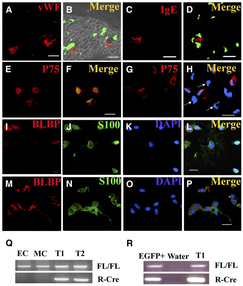
Figure 5. Only p75+ cells recombine Nf1 in Nf1 flox/flox; DhhCre mouse nerves and tumors.
EGFP expression (B, D) in sections from adult Nf1 flox/flox; DhhCre; EGFP sciatic nerves, cryo-sectioned and stained with anti-von Willebrand factor to mark endothelial cells (red, A), or anti IgE Fc receptor to mark mast cells (red, C). In B and D show merged images of EGFP fluorescence and immunostains. FACS sorted EGFP+ (E, F) and EGFP- (G, H) cytospun cells were immunostained with anti-p75 antibodies (E, G). All EGFP+ cells are p75+, but some EGFP-are p75+ while other are p75-negative cells (H, white arrows). FACS sorted EGFP+ cells from P1/P2 sciatic nerves (I–L) or tumor were stained with Blbp ( I, M, red) and S100β (J, N, green). Bars, A–P: 13 μm. FACS sorted cells from P1 sciatic nerve or tumor were analyzed for Cre mediated recombination of Nf1 by PCR (Q, R). Q, Endothelial cells (CD31+/CD45−; EC) or mast cells (c-kit+/IgE-FcR+; MC) from dissociated tumor cells did not recombine Nf1. DNA from cells from two Nf1 flox/flox; DhhCre; EGFP tumors (T1, T2) show recombination. Nf1fl allele (FL/FL), recombined Nf1 allele (R-Cre). R, postnatal day 1 EGFP+ cells (EGFP+) showed Nf1 recombination, as did tumor DNA (T1). Water is shown as a negative control.

