
Figure 4. Hb is a new substrate of MAPK in the embryo.
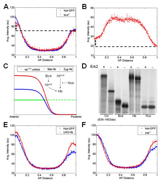
(A) Comparison of the dpERK gradients in wild type (histone-GFP) embryos and embryos from bcd null mothers. Average dpERK gradients for each genotype (N~15) are plotted with SE indicated. Although the anterior levels of dpERK in bcd null embryos are lower than that in wild type, the gradient is still asymmetric along the AP axis (arrow).
(B) Averaged AP gradient of nuclear Cic at cell cycle 14 obtained from 27 embryos laid by bcd null flies. Similar to the dpERK gradient, the Cic pattern is still asymmetric along the AP axis (arrow).
(C) Simplified description of Hb regulation in a wild type embryo. Maternal hb mRNA is deposited uniformly throughout the embryo (green), but its translation is repressed in posterior half by Nos (dashed green), resulting in anterior gradient of maternal Hb protein (blue). At the anterior half of the embryo, Bcd activates zygotic hb transcription which further adds to the pre-existing Hb gradient (red). Note that even in the absence of Bcd, Hb protein is still present in anterior gradient (blue).
(D) Radiolabelled Hb migrates with slower mobility on SDS-PAGE following incubation in the presence (+), but not in the absence (−), of activated ERK2, indicating that Hb is phosphorylated by MAPK. Similar mobility shift is observed for known targets of MAPK such as Cic and Bcd, but not for Runt. Note that Hb contains several consensus MAPK phosphorylation sites.
(E) Ubiquitous expression of hb using a maternal GAL4 driver leads to increase in MAPK phosphorylation both at the anterior and posterior poles of the embryo (t-test: pAnterior = 0.03, pPosterior = 0.01); average dpERK gradients for each genotype (N~25-30) plotted with SE indicated.
(F) Quantifying the dpERK gradient in embryos mutant for osk shows an increase in MAPK phosphorylation at the posterior region. In osk null embryos, maternal hb mRNA is translated at the posterior of the embryo. Average dpERK gradients for each genotype (N~25-30) are plotted with SE indicated.