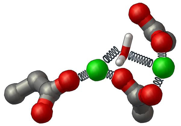
Fig. 3.
Diagram of the NMR-type restraints applied to the DDE motif, the two magnesium ions, and the bridging water between the magnesiums during Molecular Dynamics. The side-chains of the DDE motif are shown as ball-and-sticks, the magnesium ions are displayed as CPK (at reduced size to enhance clarity), and the restrained water molecule is shown as thin sticks. The restraints applied to the magnesium-oxygen interactions during the energy minimization calculations and the subsequent MD simulations are depicted as metal springs. Each spring represents a separate Mg-O restraint that was applied during the creation of these dynamic models of HIV integrase.

