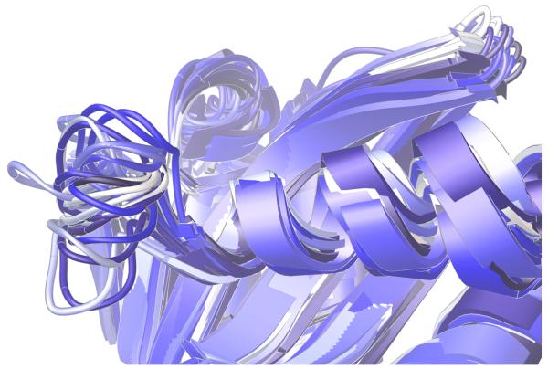
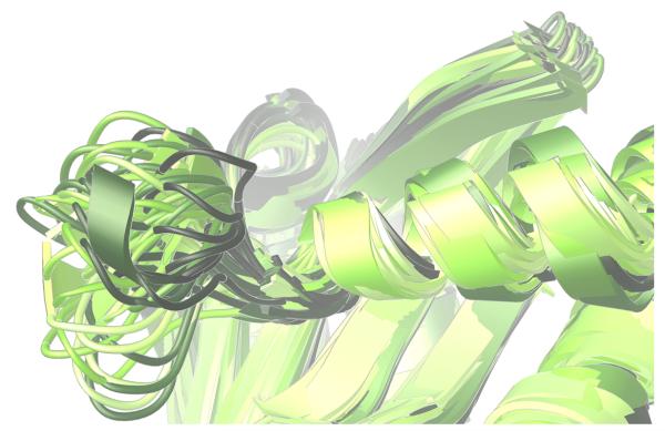
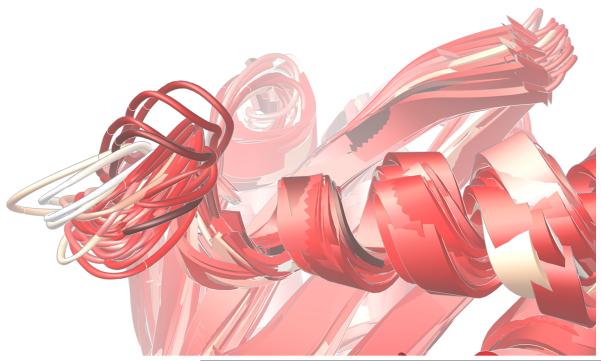
Fig. 4.
Significant differences were displayed in the dynamics of the critical 140s loop during the MD simulations of the wild type, the E92Q/N155H mutant, and the G140S/Q148H mutant of the catalytic domain of HIV integrase. Ribbon diagrams of the QR Factorization-derived ensembles extracted from MD simulations of these three variants at a QH2 = 0.89 are presented. The 140s loop is located at the left side of each image. 27 snapshots from the wild type’s MD simulation are displayed in panel a. 51 conformations of the E92Q/N155H mutant are displayed in panel b, and 47 target conformations of the G140S/Q148H mutant are presented in panel c. The darkest snapshot in each panel corresponds to the beginning of each MD simulation, and the ribbon diagrams get progressively lighter towards the end of each MD simulation. The snapshots were superimposed using the backbone atoms of the DDE motifs.


