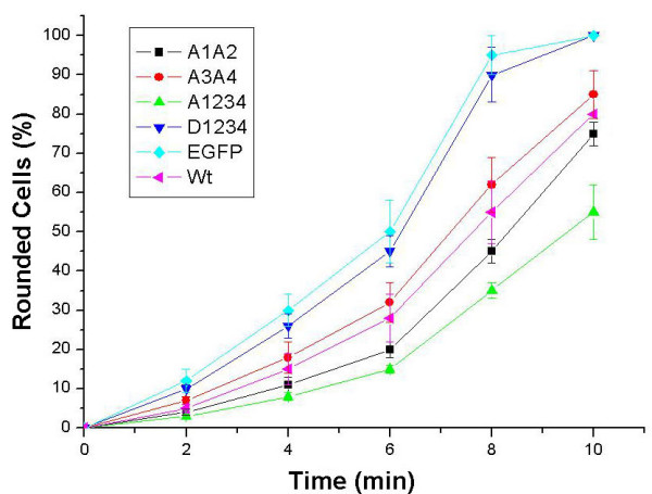
Figure 5.

Detachment of transfected A7r5 cells upon trypsinization. A1A2- (squares), A3A4- (circles), A1234- (triangles), D1234- (inverse triangles), EGFP- (diamonds), wtCaD- (turned triangles) transfected cells were plated on 60 mm dishes. Cells from each plate were trypsinized and monitored under the phase-contrast and fluorescence microscope for time-dependent retraction, rounding and detachment. Percentages of round cells at 2, 4, 6, 8 and 10 min were plotted for each type of cells. Each point was an average of 6 independent measurements; error bars represent standard deviations.
