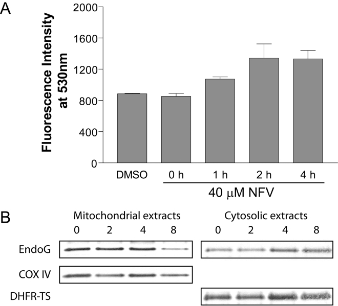
Figure 5. Loss of mitochondrial potential and translocation of EndoG in NFV-treated L. donovani amastigotes.
(A) L. donovani axenic amastigotes were exposed to the diluent or NFV (40 µM) for the indicated time periods. Mitochondrial membrane potential (Δψm) was assessed through the use of the JC-1 Mitochondrial Membrane Potential Detection Kit. Data are expressed as mean ± SD of three independent experiments. (B) Western blot analysis of the mitochondrial and cytosolic fractions obtained from NFV-treated amastigotes at different intervals. Anti-EndoG immunoblots of cytosolic and mitochondrial fractions are shown, along with the loading controls for mitochondria (i.e. COX IV) and cytosol (i.e. DHFR-TS), respectively.

