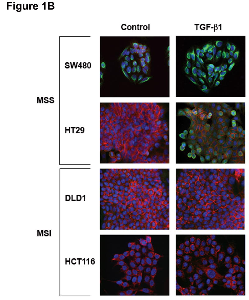
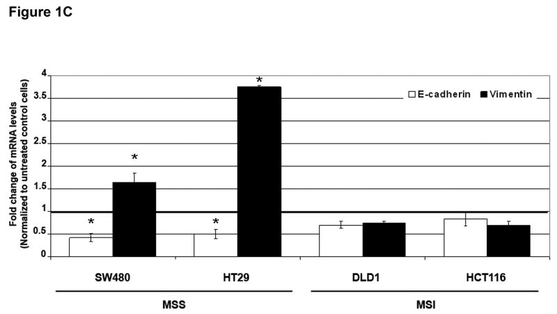
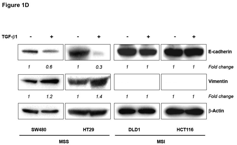
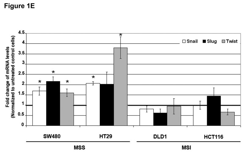
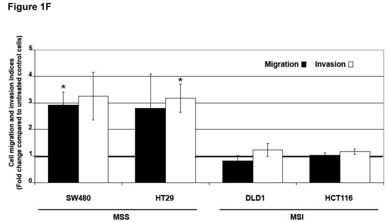
Figure 1. TGF-β 1 induces EMT in MSS but not MSI cell lines with a mutant TGFBR2.
A) Phase-contrast photomicrographs and B) immunofluorescence images of control cells and cells treated with 5 ng/mL TGF-β1 for 48 hours. In B), cells were immunostained with antibodies to E-cadherin (red) and vimentin (green) and then stained with DAPI to detect nuclei (blue). C) Relative mRNA levels of E-cadherin and vimentin in untreated cells and cells treated with 10 ng/mL TGF-β1 for 48 hours. The control values have been normalized to 1, and the data are expressed as fold change in treated cells. Columns, average of at least three independent experiments; bars, SEM. *, P<0.05 as compared to control cells. D) Western blot analysis of E-cadherin and vimentin expression in total lysates of untreated cells and cells treated with 10 ng/mL TGF-β1 for 48 hours. Densitometry values are expressed as fold change compared with control values normalized to 1. E) Relative mRNA levels of Snail, Slug and Twist, in untreated cells and cells treated with 10 ng/mL TGF-β1 for 48 hours. The control values have been normalized to 1, and the data are expressed as fold change in treated cells. Columns, average of at least three independent experiments; bars, SEM. *, P<0.05 as compared to control cells. F) Migratory and invasive behavior of untreated cells and cells treated with 5 ng/mL TGF-β1 for 48 hours. The control values have been normalized to 1, and the data are expressed as fold change in treated cells. Columns, average of at least three independent experiments; bars, SEM. *, P<0.05 as compared to control cells.


