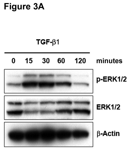
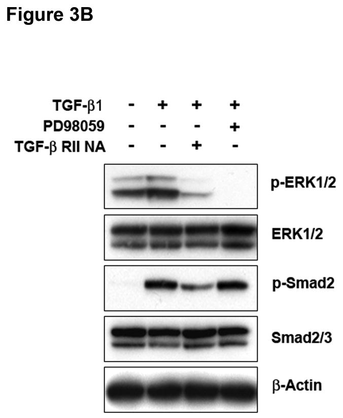
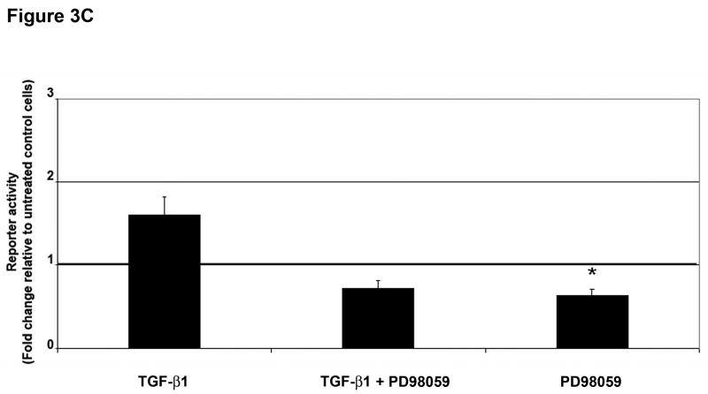
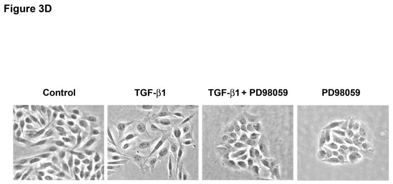
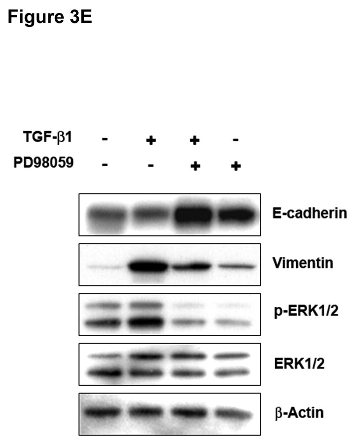
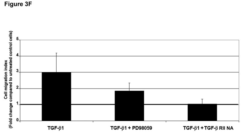
Figure 3. ERK activation can regulate TGF-β 1-induced phenotypic changes.
A) SW480 cells were treated with 5 ng/mL TGF-β1 and whole cell extracts were analyzed for the indicated primary antibodies. B) Total lysates of untreated SW480 cells, cells treated for 2 hours with 5 ng/mL TGF-β1, or cells pretreated for 60 minutes with 50 μM PD98059 or 10 μg/mL TGF-β RII neutralizing antibody were analyzed by Western blotting for the indicated primary antibodies. C) Cells were transfected with p3TP-Lux and pRL/CMV constructs and treated with 10 ng/mL TGF-β1 alone or after pretreatment with 50 μM PD98059 for 18 hours. Luciferase activity was then measured. The control values are normalized to 1, and the data are expressed as fold change in treated cells. Columns, average of at least three independent experiments; bars, SEM. *, P<0.05 as compared to control cells. D) Phase-contrast photomicrographs of SW480 cells before (control) and after treatment with 5 ng/mL TGF-β1 alone or after pretreatment with 50 μM PD98059 for 48 hours. E) The levels of E-cadherin and vimentin expression were determined by Western blot analysis in whole cell lysates from untreated SW480 cells or cells treated with 10 ng/mL TGF-β1 for 48 hours alone or after 60 minutes with 50 μM PD98059. F) Tumor cell migration was evaluated in untreated SW480 cells or cells treated with 5 ng/mL TGF-β1 alone or after 60 minutes with 50 μM PD98059 or 10 μg/mL TGF-β RII neutralizing antibody. The control values are normalized to 1, and the data are expressed as fold change in treated cells. Columns, average of two independent experiments; bars, SEM.

