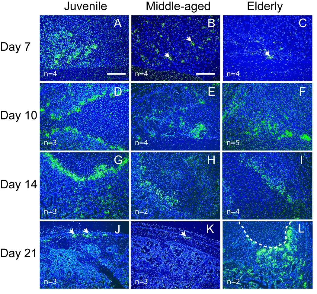
Fig. 5.
Expression of Mmp-9 detected by in situ hybridization. (A) At 7 days after fracture, robust Mmp-9 expression (green) is evident at the front of endochondral ossification in juvenile mice. (B) Weak Mmp-9 expression (arrows) is observed in middle-aged and (C) elderly mice. (D–F) At 10 days and (G–I) 14 days after fracture, strong Mmp-9 expression is present in fracture calluses of all three age groups. (J) By 21 days, Mmp-9 expression (arrows) is limited to the periosteum in juvenile and (K) middle-aged mice, (L) but a large amount of cartilage (outlined) and Mmp-9 expression is still present within the calluses of the elderly. Scale bar: A, D–L = 200 µm, B and C = 100 µm.

