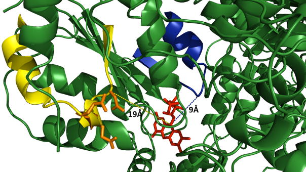
Fig. 1.
Proposed docking sites for protein redox partners and CPR based on the X-ray crystallographic structure [49]. Loops potentially mediating protein-protein interactions are shown relative to the electron donating FMN moiety. FMN, red; α helix F-β sheet 5 loop, blue; β sheet 2-α helix C loop, yellow; mutated acidic residues, orange. Molecular graphics were generated by PYMOL (http://pymol.sourceforge.net).

