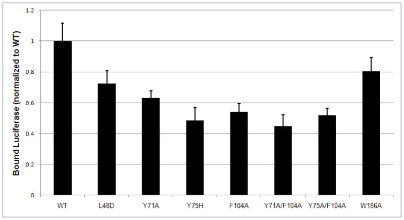
Fig. 5.
P58(IPK) binds the misfolded proteins via the conserved hydrophobic patch within the groove of domain I. The misfolded protein binding assay was performed as in Fig. 3 using Luciferase as the model protein. P58(IPK) TPR fragment (P58 TPR WT, residue 33–393) effectively interacts with the misfolded Luciferase. The mutations for the hydrophobic residues within the groove of domain I significantly reduced the binding ability of P58(IPK) TPR fragment to the misfolded Luciferase. The mutation (W186A) at the junction of domain I and II does not affect the binding.

