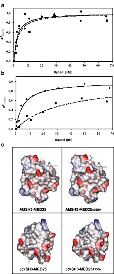
Fig. 3.

Affinity test results for MED25 binding to Abl SH3 (a) and Lck SH3 (b). Fluorescence titration experiments were performed for wild-type (filled square, dotted line) and mutant MED25 peptides carrying an A335V point mutation (filled circle, solid line). Curves were fitted according to the equation given in methods. c Models of MED25 in complex with Abl SH3 (top) and Lck SH3 (bottom) visualizing the effect of the A335V mutation. The SH3 domains are shown in space-filled representation and the acidic and basic surface patches are colored in red and blue, respectively. White surface patches correspond to nonpolar regions. The MED25 binding region is shown as tube and residue 335 is shown in stick presentation
