Figure 3.
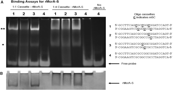
Binding Assays for rMcrA-S. (A) Assays of electrophoretic mobility shifts with different molar ratios (1 : 1 and 1 : 4—DNA:protein) of rMcrA-S binding unmethylated and symmetrically methylated (m5CpG) containing 24-bp double-stranded oligonucleotides: 1 contains three m5C including one in an HpaII site; 2 contains a single m5C in a ‘three–fourth HpaII site’; 3 contains a single m5C in an HpaII site; 4 has no m5Cs. Molar ratios are based on monomeric McrA (16). Asterisks indicate the positions of McrA-S/DNA complexes. (B) Coomassie Blue staining of EMSA (A) gel.

