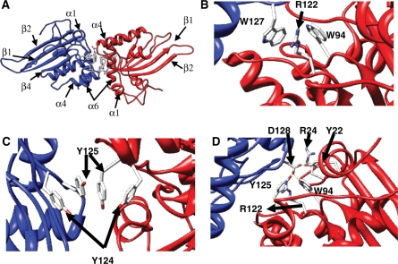
Figure 1.
Homology model of the Apobec3G N-terminal deaminase domain. A dimer of two N-terminal domains (red and blue), interacting via the 122–127 motif is shown. (A) Overview with indication of secondary structure elements. (B–D) Detail of selected sidechain interactions between the different monomers.

