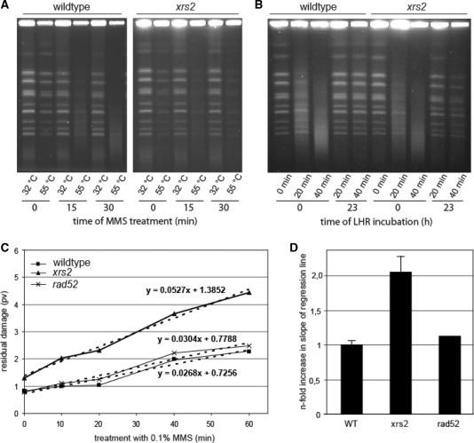
Figure 2.
Analysis of the repair of heat-labile sites. (A) Induction of chromosomal degradation in wildtype and the xrs2 mutant visualized by pulsed-field gel electrophoresis. Cells were treated for 0, 15 and 30 min with 0.1% MMS. During preparation of chromosomal DNA, the incubation with proteinase K was carried out at 32°C (heat-labile sites remain stable) as well as 55°C (heat-labile sites are converted into DSB). (B) Analysis of chromosomal degradation (proteinase K incubation at 55°C) immediately after treatment for 0, 20 and 40 min with 0.1% MMS as well as after 23 h incubation of the cells under LHR conditions. (C) Residual DNA fragmentation plotted against the time of MMS treatment. The degree of residual chromosomal degradation was quantified using the software program Geltool and is shown as profile value (pv). Linear equations are calculated from regression lines (dashed lines). One representative experiment is shown. (D) Slope of regression lines obtained from residual chromosomal degradations from the xrs2 and rad52 mutant relative to wildtype. Mean values from three independent experiments for the xrs2 mutant and for wildtype are shown. The rad52 mutant was analyzed once.

