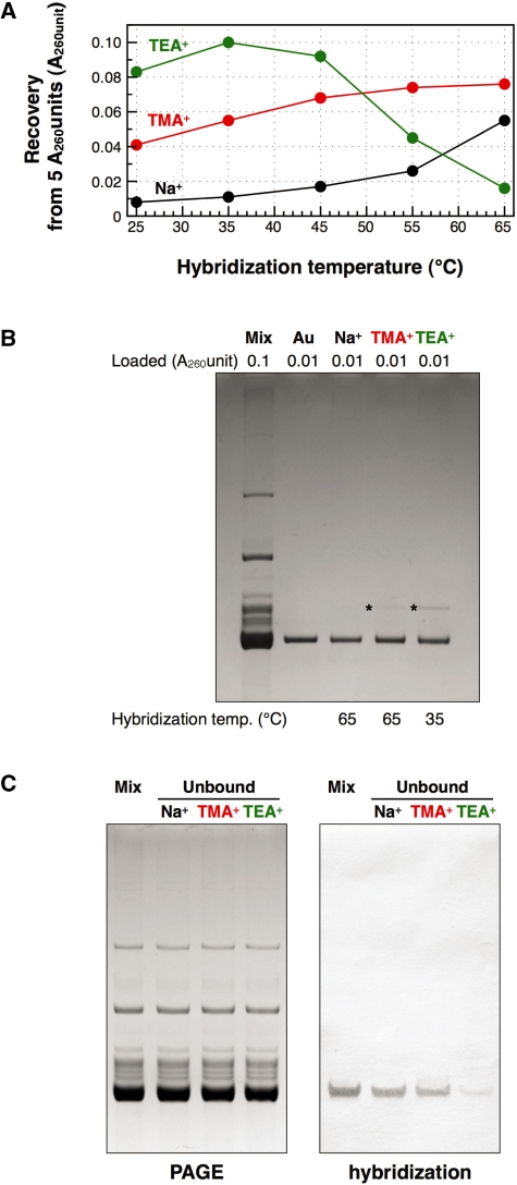
Figure 5.
Quantities and qualities of the recovered E. coli
 under various hybridization conditions. (A) Recovered amounts of the tRNA from 5 A260 units of unfractionated E. coli tRNAs at various hybridization temperatures. (B) Analysis by 10% PAGE containing 7 M urea. Lane mix: 0.1 A260 units of unfractionated E. coli tRNAs. Lane Au: 0.01 A260 units of the authentic E. coli
 . Lanes Na+, TMA+ and TEA+: 0.01 A260 units of the recovered E. coli
 with hybridization buffer Na+ (0.9 M NaCl, hybridized at 65°C), hybridization buffer TMA+ (TMA-Cl, hybridized at 65°C) and hybridization buffer TEA+ (TEA-Cl, hybridized at 35°C), respectively. The gel was stained with methylene blue. The asterisk shows a contaminant band found in the recovered E. coli
 . (C) Northern hybridization to detect E. coli
 in the tRNA fractions unbound to the resin. (Left) Lane mix: 0.1 A260 units of unfractionated E. coli tRNAs. Lanes Na+, TMA+ and TEA+: 0.1 A260 units of the tRNA fractions unbound to the resin using hybridization buffer Na+ (hybridized at 65°C), hybridization buffer TMA+ (hybridized at 65°C) and hybridization buffer TEA+ (hybridized at 35°C), respectively. The gel was stained with methylene blue. (Right) Northern hybridization analysis with Oligo-EfMet. Biotins on the nylon membrane were detected with streptavidin, bitotinyl alkaline phosphatase and BCIP/NBT substrate.

