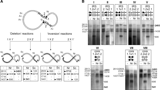
Figure 7.
Analysis of HJ intermediates formed at different variants of IRS. HJ products that accumulated in recombination reactions performed with different IRS substrates (see the ‘Materials and Methods’ section) were cut with AvaII (Av), gel purified, and 5′-labelled with 32P (asterisk). The resulting χ-forms were left untreated (−) or recut with NruI (Nr) or ScaI (Sc) and run on DNA denaturing gels. (A) Different patterns of radio-labelled single-stranded DNA fragments are expected depending on whether the HJ is formed by the IR1-bound TnpI subunits (1 × 1′), the IR2-bound subunits (2 × 2′) or opposite combinations of IR1- and IR2-bound subunits (1 × 2′ and 2 × 1′, respectively). Sizes (in nt) of the diagnostic fragments obtained for the different possibilities of strand exchange are reported in the corresponding tables. One fragment (in bold) is specific for each possibility and the corresponding DNA strand is shown as a thick line in the cartoons showing the different HJ isoforms. (B) Bands patterns obtained for the different substrates after autoradiography of the gels. For each group of substrates, sizes of the initial χ-form fragments are indicated on the left, and the new fragments that were generated after NruI and ScaI cleavage are shown on the right. All the DR1–DR2-constraining substrates (panels I–V) show a unique pattern of bands corresponding to the 1 × 1′ exchange, irrespective of the nature and orientation of the core sites. The pattern obtained for the pCore1.2 × 1.2 substrate (panel VI) is consistent with strand exchange occurring at both IR1 and IR2. HJ formation at the symmetrical Core1.1 and Core2.2 sites (panel VII and VIII) occurred with all possibilities of strand exchange.

