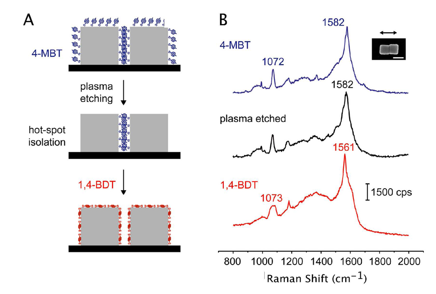
Figure 7.
Probing the hot spot within a dimer of silver nanocubes. (A) Schematic of the approach employed for selectively probing the hot spot region formed between a pair of silver nanocubes. The dimer was functionalized with 4-methyl benzenethiol (4-MBT) and then exposed to plasma etching to remove the adsorbed 4-MBT molecules. In this case, only the 4-MBT molecules outside the hot spot region (i.e., outside the two touching faces) were removed during the plasma etching. The nanocube dimer was then immersed in a 1,4-benzenedithiol (1,4-BDT) solution, resulting in the complete replacement of 4-MBT by 1,4-BDT over its entire surface. (B) The corresponding SERS spectra, where the middle spectrum represents the SERS signals from molecules in the hot spot region only. The scale bar represents 100 nm. Copyright (2009) Wiley-VCH. Reprinted with permission.

