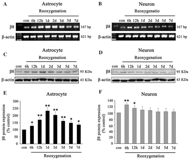
Fig. 2.
The effects of HI on β8 expression in cultured astrocytes and neurons. β8 mRNA expression was increased at 6 h after reoxygenation, peaked at 1 day, then slowly decreased but still maintained at high level at 7 days in astrocytes compared with controls (a). HI also resulted in an increase of β8 mRNA expression in neurons at 6 h, but declined at 12 h and returned to baseline within 2 days after reoxygenation (b). Analysis showed that the β8 protein was increased at 6 h, and maintained for at least 7 days after reoxygenation in astrocytes (c), but slightly and transiently increased in neurons at 6 h after reoxygenation and quickly returned to baseline within 1 day (d). Quantification of β8 expression in astrocytes and neurons, respectively, in HI groups and normoxic controls (e, f). Results were normalized to controls and represented as mean ± SD. For each column, N = 4, * P < 0.05, ** P < 0.01 versus control

