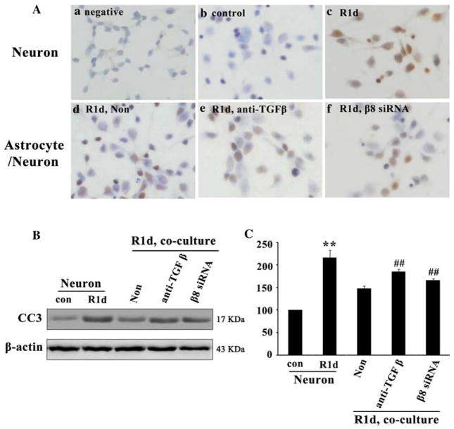
Fig. 5.
Integrin β 8/TGF- β 1 signaling pathway protected neurons from apoptosis after OGD. TUNEL positive cells expressed stronger in neurons at 1 day after reoxygenation (Ac, R1d) compared with the neurons in normoxic controls (Ab, con) and in negative controls (Aa). The expression of TUNEL positive cells were significantly decreased while co-cultured with astrocytes (Ad, Non). However, after blocking TGF-β 1 or blocking β 8, the number of TUNEL positive cells was increased (Ae, Af). Western blot analysis showed that CC3 expression was significantly increased at 1 day after reoxygenation in neurons cultured alone (B, R1d) compared with the normoxic controls. CC3 expression was significantly decreased while co-cultured with astrocytes (B, Non), but was rescued while blocking TGF-β (B, anti-TGF β) or β 8 (B, β8 siRNA). Quantification of CC3 expression in neurons cultured alone and neurons co-cultured with astrocytes (C). Results were normalized to normoxic controls and represented as mean ± SD. For each column, N = 4, **P < 0.01 versus con & Non, ## P < 0.01 versus Non

