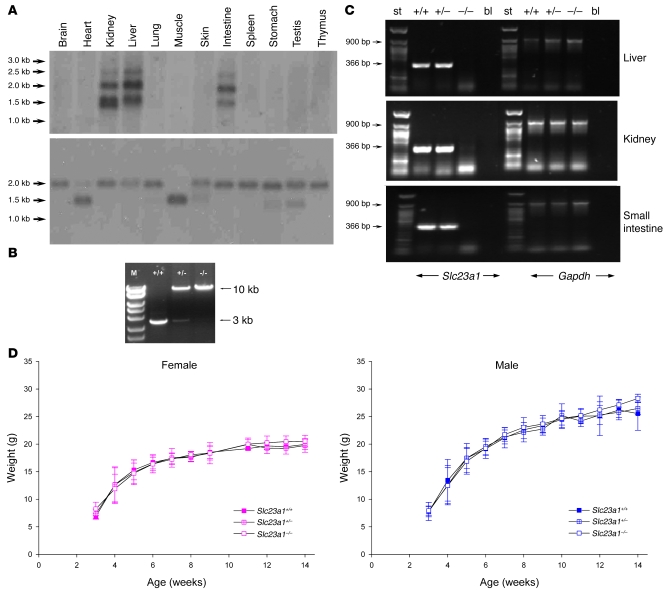
Figure 1. Design, preparation, and confirmation of Slc23a1–/– mice.
(A) Northern blot analysis of mouse Slc23a1 gene expression. Multitissue mouse Northern blot panel probed with [α32P]-d-CTP–labeled mouse Slc23a1 cDNA (top), normalized to β-actin gene expression (bottom). (B) Genomic DNA PCR analyses. DNA obtained from Slc23a1+/+, Slc23a1+/–, and Slc23a1–/– littermates was analyzed by PCR. The wild-type allele was predicted to be a 3-kb fragment; the deletion allele was predicted to be a 10-kb fragment. M, DNA marker standards. (C) RT-PCR analysis of Slc23a1 gene expression in progeny from heterozygous crosses. Gene expression was assessed in liver, kidney, and small intestine from Slc23a1+/+, Slc23a1+/–, and Slc23a1–/– mice. A 366-bp fragment was amplified by RT-PCR in Slc23a1+/+ and Slc23a1+/– RNA, but not in RNA isolated from Slc23a1–/– mice. As an internal control, gene expression of Gapdh was assessed by amplifying a 900-bp GAPDH PCR product in all tissues analyzed. bl, blank control; st, DNA standards. (D) Body weight as a function of time in Slc23a1+/+, Slc23a1+/–, and Slc23a1–/– mice. Female (n = 3–19) and male (n = 4–29) littermates were weighed at weaning (3 weeks) and weekly thereafter. P = NS.

