Figure 2.
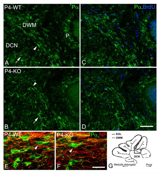
Distribution and proliferation of oligodendrocyte progenitors at the 4th postnatal day. In contrast to stellate-shaped (arrows) PDGFRα-immunoreactive (Pα) OPCs, which were observed mainly in deep cerebellar nuclei (DCN), longitudinally-oriented OPCs (arrowheads) were seen mainly in the developing white matter (DWM) of wild type (A, C, E; WT) and NG2-null (B, D, F; KO) pups at the 4th postnatal day (P4). Fewer longitudinally-oriented OPCs were distributed between neurofilament-positive cerebellar axons (NF, red; D - dapi, blue) in the DWM of NG2-null animals (F). In contrast to the DCN, BrdU/PDGFRα-positive OPCs were more abundant in the DWM of wild type (C) and NG2-null (D) pups at the 4th postnatal day. Some BrdU/PDGFRα-positive OPCs were also observed in the Purkinje cell layer (P). The schematic drawing (G) depicts the developing white matter (DWM), deep cerebellar nuclei (DCN) and the external granular layer (EGL) at the level of the superior cerebellar peduncle. The solid rectangle (G) indicates the region shown in other images (A-D). Scale bars: 20 μm (E, F), 50 μm (A-D).

