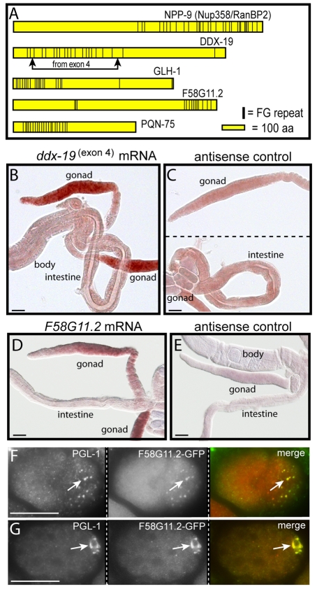
Fig. 9.
FG-repeat proteins in P granules. (A) Diagram showing FG repeats (vertical bars) in the nucleoporin NPP-9 and in four additional C. elegans proteins encoded by gonad-enriched mRNAs. (B,C) In situ hybridization for sense (B) and antisense (C) ddx-19(exon4) mRNA. (D,E) In situ hybridization for sense (D) and antisense (E) f58g11.2 mRNA. (F)Two-cell embryo showing colocalization of F58G11.2::GFP with cytoplasmic P granules (arrow). (G) Fifty-cell embryo showing colocalization of F58G11.2::GFP with P granules (arrow). Scale bars: 25 μm.

