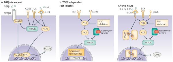
Figure 4. Mechanisms of FOXP3 induction in naïve T cells.
a | Stimulation of FOXP3− T cells through TCR and CD28 in the presence of TGFβ promotes expression of the FOXP3 gene through the cooperation of NFAT and mothers against decapentaplegic homologue 3 (SMAD3). This process is counteracted by mTOR activation, which explains the increased expression of FOXP3 when stimulation takes place in the presence of rapamycin (which inhibits mTOR activation). b | Limited TCR–CD28 stimulation (<18 hours) promotes a PI3K–AKT–mTOR-mediated re-organization of chromatin that includes increased accessibility to the FOXP3 gene. Prolonged TCR/CD28 stimulation prevents, again through activation of the PI3K–AKT–mTOR pathway, the expression of FOXP3 which would otherwise probably be induced by signal transducer and activator of transcription (STAT)5-activating cytokines generated during the initial stimulation. This two-step model rationalizes the opposing effects of rapamycin administration during T-cell activation that have been observed, which probably depend on the timing of mTOR inhibition.

