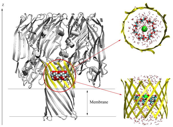
Figure 1.
Molecular graphics view of the GSBP simulation system for (M113N)7 channel with βCD adapter inside. The full protein is shown in cartoon style (grey), while the inner GSBP simulation region is highlighted in yellow. Channel axis is along the z-axis. Implicit membrane is centered at z = 0 Å with thickness of 25 Å. The reduced system for GSBP is zoomed in and shown in top view and side view. The residues in the inner region, yellow ribbons; βCD, VDW model in whole channel and licorice model in zoom-in figure; Cl−, green ball; TIP3 water, stick in atom type colors.

